Vp Shunt Skull X Ray

Fractured Vp Shunt Frontal Radiograph Of The Cervical Spine Edge Enhanced Shows A Fracture Of The Ventriculo Peritoneal Shunt Dr Vp Shunt Pet Ct Radiography

My Granddaughters Xray Due To Hydrocephalus Granddaughter Awareness X Ray

Vp Shunt Tubes For Hydrocephalus Google Search Vp Shunt Lins Looking Up

Vp Shunt System Vp Shunt Brain Surgery Epilepsy

Diagnosis Veterinary Radiology Animal Medicine Vet Tech Student

Journal Of Perinatology Figure 2 For Article Trisomy 14 Mosaicism A Case Report And Review Of The Literature Genetic Disorders Mri Neurology

See ventriculoperitoneal shunt complications.
Vp shunt skull x ray. Patients may have negative ct findings but positive x ray findings 2. Ensuring clear visualization of the shunt in the skull and its path down the cervical region. 8 views ap and lateral abdomen ap and lateral chest ap and lateral c spine ap and lateral skull.
The programmable csf shunt valve has become an important tool in hydrocephalus treatment particularly in the nph population and in pediatric patients with complex hydrocephalus. The shunt series is a set of radiographic images performed to assess the location and integrity of a ventriculoperitoneal shunt. Usually a simple lateral skull x ray helps to identify the type of valve and it s adjustment.
Shunt series ap lateral skull and chest abdominal x ray as a baseline for future comparison some surgeons obtain these films immediately post op in case some immediate revision is indicated e g. Ventriculoperitoneal vp shunts are a device used to shunt cerebrospinal fluid in the treatment of hydrocephalus. The purpose of this study is to provide a single reference for the identification of programmable shunt valves and the interpretation of programmable shunt valve settings.
A ventriculoperitoneal vp shunt is a medical device that relieves pressure on the brain caused by fluid accumulation. Shunt series for vp shunt x ray guideline. Reviewed 2016 amr.
The verification of shunt adjustment so called opening pressure and also the primary identification of the implanted valve type can be a problem in daily practice. Frontal radiograph of the cervical spine edge enhanced shows a fracture of the ventriculo peritoneal shunt drainage tube black and white arrows with caudal retraction of the distal fragment. If the shunt is somewhat superimposed on.
Ventricular catheter tip in temporal horn. The external portion of the catheter is connected to a valve that regulates the flow of csf based on a preset pressure.

Pin On Excalibur Healthcare S Imaging Teleradiology Pins

Connatal Cyst Mri Nuclear Medicine Cns Mri
Brain Tumors Children S Hospital Of Wisconsin Child Life Specialist Child Life Childrens Hospital

Pectus Excavatum Nuss Pectus Excavatum And Nuss Procedure Pectus Excavatum Pediatric Surgery Surgery

Progressive Brain Changes In Patients With Chronic Fatigue Syndrome Are Our Brains Starved Of Oxygen Latter

Common Endoscopic Functional Injury Sinus Site Surgery A Common Site Of Injury During Functional Endos In 2020 Medical Therapy Sinus Surgery Cerebrospinal Fluid
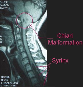
Image From Http Asap Org Wp Content Uploads 2009 10 Mri Jpg Chiari Chiari Malformation Occipital Neuralgia

Cranio Cervical Syndrome Ccs Symposium April 6 2013 Raymond V Damadian Md Cervical Spine Health Syndrome

Ct Scan Of Successful Chiari Decompression 3in By 2in Skull Removal In Tact Tonsils C1 Laminectomy Duraplasty Wit Chiari Spinal Fluid Chiari Malformation

Pin By محمد رزق On Radiologija Radiology Medical Imaging Radiography

Pin On Sumer S Radiology Site

Pin By Maher Zaben On Father Brain Case 2020 In 2020 Intracranial Pressure Cerebrospinal Fluid Normal Pressure

Seizures And Epilepsy Frequently Asked Questions This Or That Questions Seizures Epilepsy
