Vp Shunt Setting X Ray
Determining Settings Of Programmable Vp Shunts Uw Emergency Radiology

Programmable Csf Shunt Valves Radiographic Identification And Interpretation American Journal Of Neuroradiology
Programmable Cerebrospinal Fluid Shunt Radiology Reference Article Radiopaedia Org

Radiologic Identification Of Vp Shunt Valves And Adjustment Pediatric Neurosurgery Leipzig
Brg Reading Skull Films For Shunt Valve Settings

Codman Hakim Programmable Vp Shunt Radiology Case Radiopaedia Org

Lollis ss mamourian ac vaccaro tj duhaime a c.
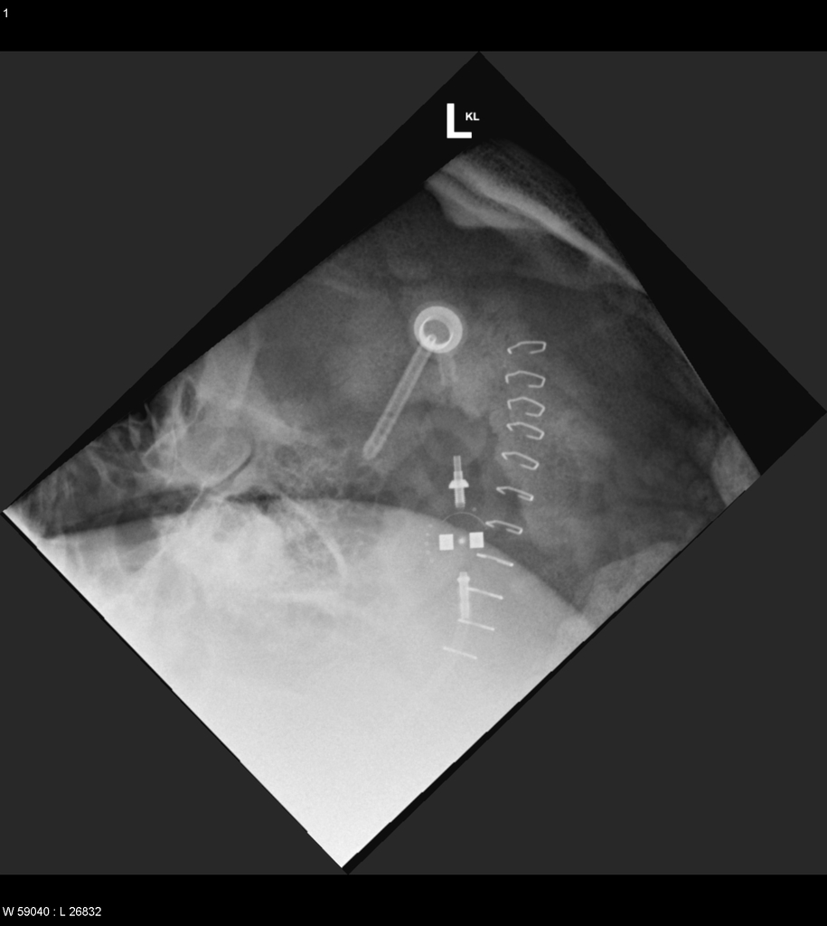
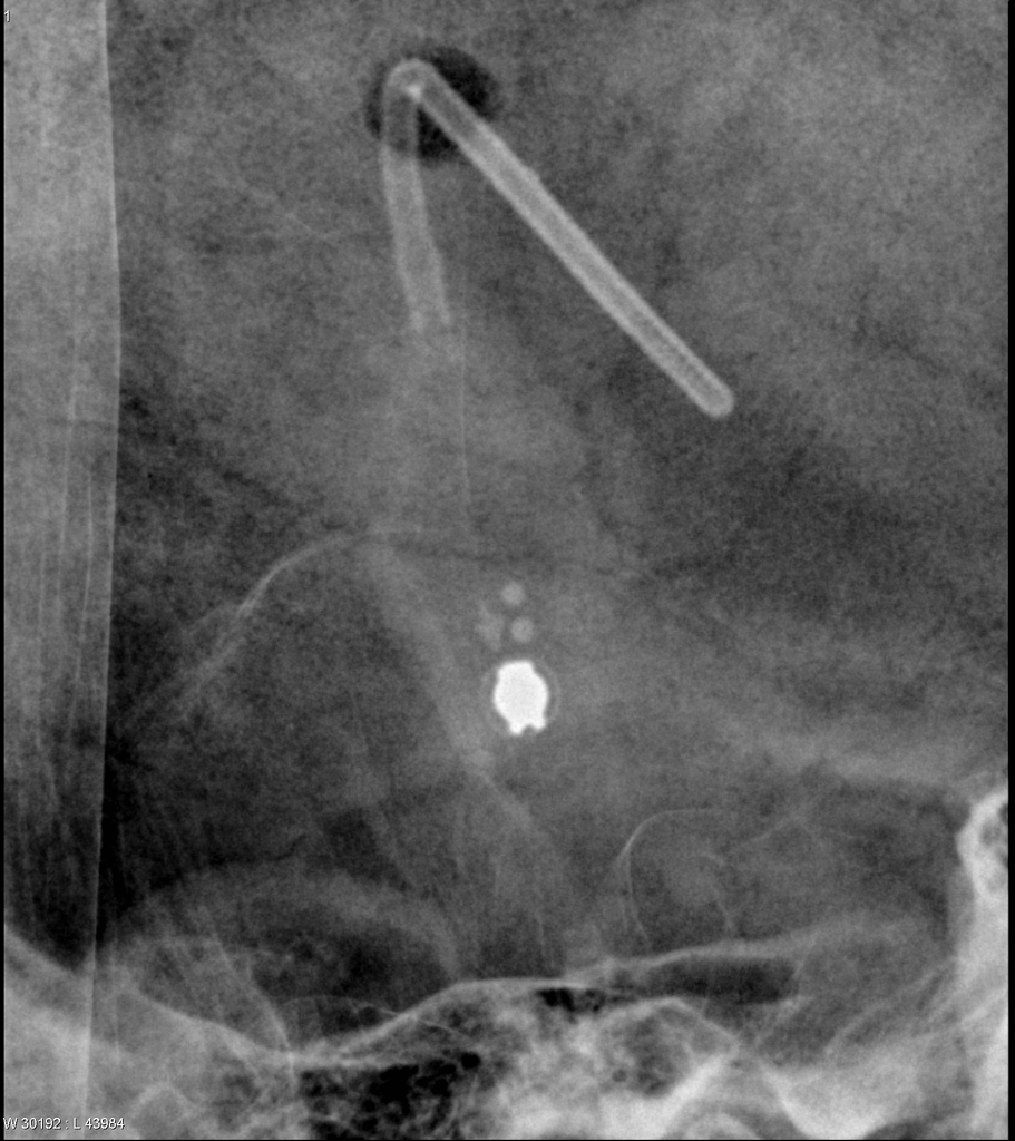
Vp shunt setting x ray. Usually a simple lateral skull x ray helps to identify the type of valve and it s adjustment. The shunt series is a set of radiographic images performed to assess the location and integrity of a ventriculoperitoneal shunt. 8 views ap and lateral abdomen ap and lateral chest ap and lateral c spine ap and lateral skull.
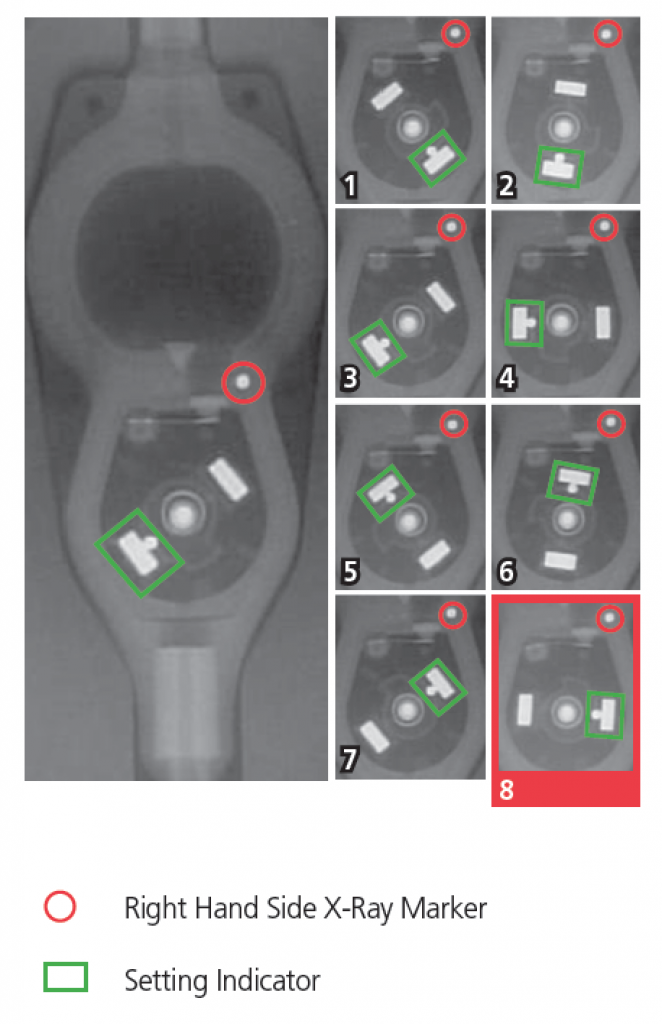
Each performance level. C setting code for aesculap miethke progav gravitational unit. Programmable cerebrospinal shunts are a type of ventriculoperitoneal shunt that can be set to different csf pressure settings.
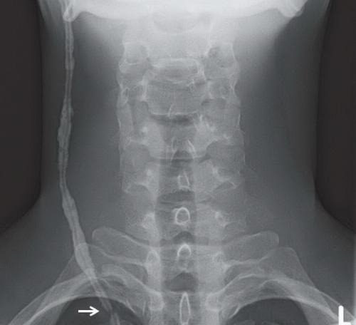
Radiographic identification and interpretation. Frontal radiograph of the cervical spine edge enhanced shows a fracture of the ventriculo peritoneal shunt drainage tube black and white arrows with caudal retraction of the distal fragment. Shunt pass is not always at hand and documents from external hospitals can be missing.
American journal of neuroradiology. C conventional radiographs of head neck and chest depict ventriculostomy catheter placed via right frontal approach white arrow a and b and normal course of distal ventriculoperitoneal shunt catheter terminating in lower abdomen arrowheads. In summary the optimal setting for the treatment of a patient with inph is a vp shunt with an opening pressure of 5 cm h 2 o and a gravitational unit shunt assistant prosa which will prevent overdrainage at 20 cm h 2 o.
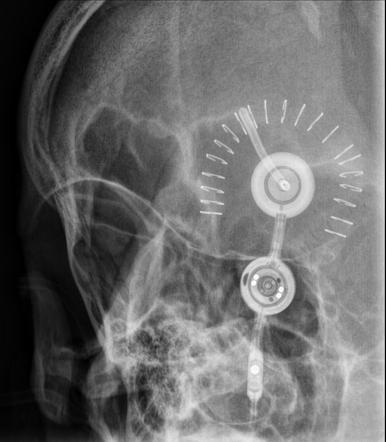
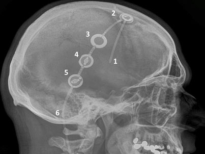
1 provides radiographic features with reference images of the settings of five common valves codman hakim programmable valve medtronic. A complete aesculap miethke progav assembly. Note redundancy of intraperitoneal catheter to allow for vertical growth of child black arrow c.
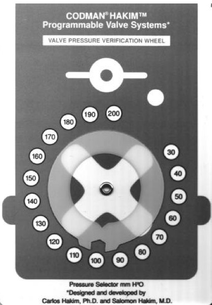
Programmable csf shunt valves. Codman certas plus x ray procedure guide brochure. The 2010 ajnr article by lollis et al.

Chest X Ray Showing Ventriculoperitoneal Shunt Tip Ascending The Download Scientific Diagram

Ommaya Ventricular Access Device And Vp Shunt Radiology Case Radiopaedia Org

Vp Shunt Disconnection Radiology Case Radiopaedia Org
Medtronic Strata Programmable Vp Shunt Radiology Case Radiopaedia Org
A Radiographic Appearance Of The Codman Hakim Programmable Valve Set Download Scientific Diagram

Abnormal Vp Shunt Series Radiology Case Radiopaedia Org
Saarland University Medical Center Shunt Implantation

Fig 3 Programmable Csf Shunt Valves Radiographic Identification And Interpretation American Journal Of Neuroradiology

Sophysa Shunt Manufacturer For Hydrocephalus Treatment
Ventriculoperitoneal Shunt Malfunction Radiology Key

Figure 7 From Imaging Of Ventricular Shunts Semantic Scholar

Ventriculoperitoneal Shunt Disruption Radiology Case Radiopaedia Org
Https Pubs Rsna Org Doi Pdf 10 1148 Radiographics 18 3 9599388