Vp Shunt Radiopaedia

Ommaya Ventricular Access Device And Vp Shunt Radiology Case Radiopaedia Org

Ventriculopleural Shunt Radiology Case Radiopaedia Org

Abnormal Vp Shunt Series Radiology Case Radiopaedia Org

Ventriculoperitoneal Shunt Disruption Radiology Case Radiopaedia Org

Vp Shunt Disconnection Radiology Case Radiopaedia Org

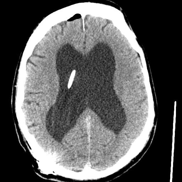
Acute Hydrocephalus From Vp Shunt Blockage Radiology Case Radiopaedia Org

Ventriculoperitoneal vp shunts are a device used to shunt cerebrospinal fluid in the treatment of hydrocephalus.
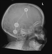
Vp shunt radiopaedia. Programmable cerebrospinal shunts are a type of ventriculoperitoneal shunt that can be set to different csf pressure settings. An example of a normal vp shunt series. The vp shunt on the right side of the neck seen best on the lateral neck skull and ap neck views appears discontinuous.
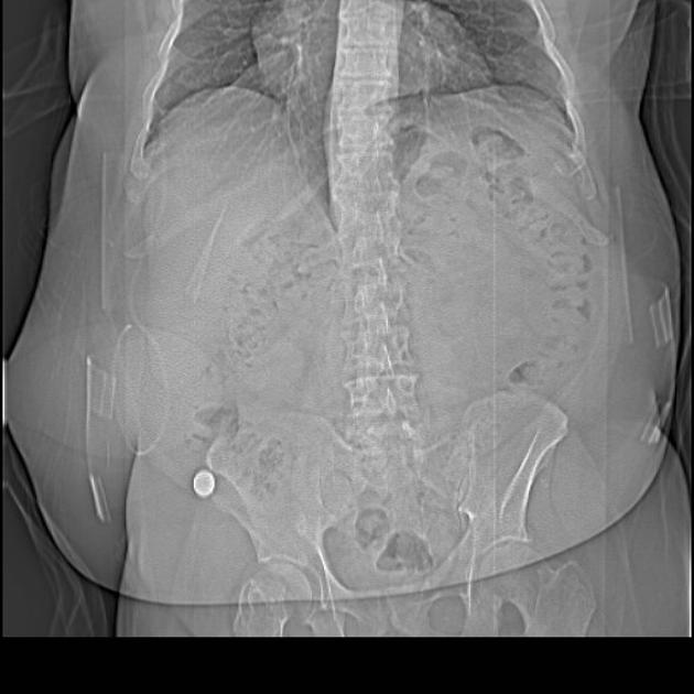
The shunt series in this patient demonstrate a vp shunt disconnection at the level of the lower neck best appreciated on the ap cervical spine view. Case contributed by radswiki. Indications departmental protocols will vary but the overall goal is to image the shunt in its entirety to assess.
Find out more. They are of particular value in normal pressure hydrocephalus and in pediatric patients. 1 provides radiographic features with reference images of the settings of five common valves codman hakim programmable valve medtronic.
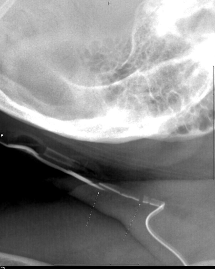
An important series of films for checking the integrity of a in situ vp shunt. The shunt series is a set of radiographic images performed to assess the location and integrity of a ventriculoperitoneal shunt. Case contributed by dr adam eid ramsey.
The shunt is not seen on the chest or abdominal wall with the remainder of the tubing coiled in the abdomen. Misplaced ventriculoperitoneal shunt catheter. Radiopaedia supporter during december and be in the running to win one of four 12 month all access passes.
The migration of a vps into both trunks of the pulmonary artery is a very rare complication. The cause for the catheter migration was a penetration of the right subclavian vein during the initial shunt placement. The 2010 ajnr article by lollis et al.
Programmable Ventriculoperitoneal Shunt Radiology Case Radiopaedia Org
Shunt Series Radiology Reference Article Radiopaedia Org
Ventriculoperitoneal Shunt Radiology Reference Article Radiopaedia Org

Normal Vp Shunt Series Radiology Case Radiopaedia Org

Normal Shunt Series Radiology Case Radiopaedia Org
Broken Vp Shunt Radiology Case Radiopaedia Org

Vp Shunt Migration Radiology Case Radiopaedia Org
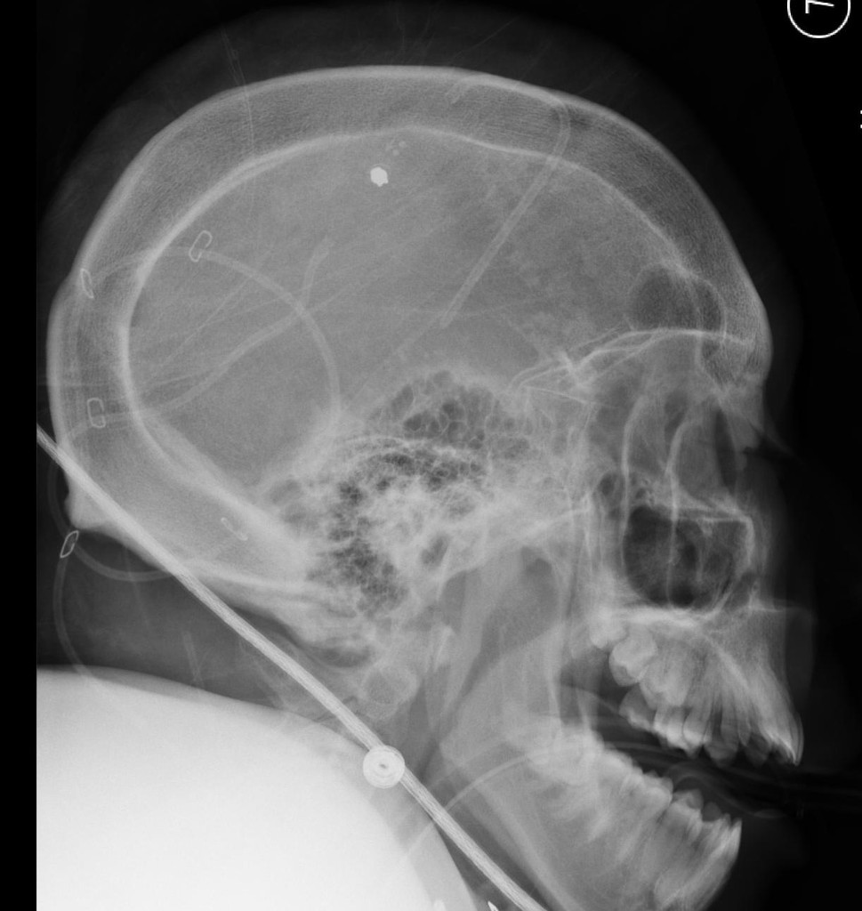
Calvarial Thickening From Chronic Ventricular Shunting Radiology Case Radiopaedia Org

Normal Vp Shunt Series Radiology Case Radiopaedia Org

Lumboperitoneal Shunt Radiology Reference Article Radiopaedia Org

Ventriculoperitoneal Shunt Radiology Reference Article Radiopaedia Org Vp Shunt Brain Surgery Radiology
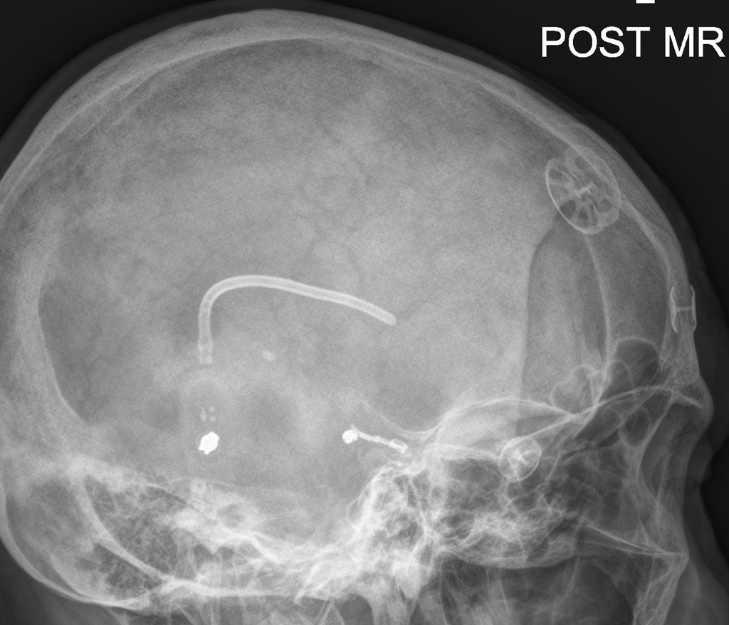
Programmable Medtronic Ventriculoperitoneal Shunt Radiology Case Radiopaedia Org

Normal Shunt Series Radiology Case Radiopaedia Org