Vp Shunt Ct Scan

Radiological Findings Before And After Vp Shunt Placement Notes A Download Scientific Diagram

Postoperative Ct Scan Of The Patient Axial View Showing Placement Of Download Scientific Diagram
Programmable Ventriculoperitoneal Shunt Radiology Case Radiopaedia Org

Brain Ct Scan Before Extraction Of Ventriculoperitoneal Shunt Download Scientific Diagram

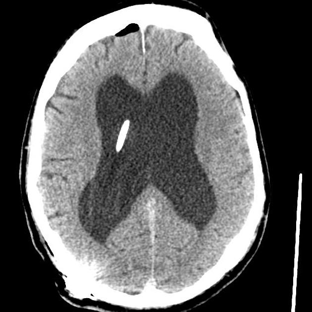
Acute Hydrocephalus From Vp Shunt Blockage Radiology Case Radiopaedia Org

Ct Scan Obtained 3 Years After Vu Shunt Placement The Left Lateral Download Scientific Diagram

Further prospective and multidisciplinary studies are needed to evaluate the reliability of limited head ct for the clinical evaluation of vp shunt malfunction.
Vp shunt ct scan. The external portion of the catheter is connected to a valve that regulates the flow of csf based on a preset pressure. In patients with ventriculoperitoneal shunts pseudocysts are caused by peritoneal adhesions or migration of the greater omentum over the shunt tip. Shunt obstructions may be confirmed with radioisotope examination or with fluoroscopically guided injection of iodinated contrast material into the shunt reservoir.
A ventriculoperitoneal shunt drains. Fluid buildup can increase brain pressure which can be harmful. Most pediatric patients with hydrocephalus are treated with ventriculoperitoneal vp shunt placement.
The purpose of a ventriculoperitoneal shunt is to remove excess fluid from a person s brain. Diagnosis of disconnection or fracture is confirmed on conventional radiographs shunt series which show entire length of shunt from skull to abdomen. Pseudocysts can also develop around ventriculopleural shunts due to adhesions caused by chronic pleural irritation.
As the name suggests a catheter is placed with its tip in the ventricle. Shunt must be pulled and have ability to move in the subcutaneous tissue. Hydrocephalus with an enlarged fourth ventricle.
Ventriculoperitoneal vp shunts are a device used to shunt cerebrospinal fluid in the treatment of hydrocephalus. However shunt malfunction is common and is usually caused by mechanical failure. Our pilot study demonstrates that utilization of limited head ct scan in the evaluation of children with suspected vp shunt malfunction is a feasible strategy for the evaluation of the ventricular size.
Diversion of csf by means of a shunt placed between the ventricular system of brain and the peritoneal cavity or right atrium may result in rapid relief of symptoms in obstructive hydrocephalus. Pseudocysts are loculated collections of csf that form around the terminal end of the catheter.

Ventriculoperitoneal Shunt Malfunction Radiology Key

Non Contrast Ct Scan Of Head Showing Evd In Situ A Followed By Right Download Scientific Diagram

A Postoperative Computed Tomography Ct Scan Of The Initial Download Scientific Diagram
Https Pubs Rsna Org Doi Pdf 10 1148 Radiographics 18 3 9599388

Ventriculoperitoneal Shunt Malfunction Malposition Misposition Misplacement Pediatric Radiology Reference Article Pediatricimaging Org Pedsimaging

Axial And Sagittal Ct Demonstrating Ventriculoperitoneal Vp Shunt Download Scientific Diagram

Shunt Systems Hydrocephalus Association

Brain Ct Scans Showing Normal Ventricles After Ventriculoperitoneal Download Scientific Diagram

Pdf Detection Of Ventricular Shunt Malfunction In The Ed Relative Utility Of Radiography Ct And Nuclear Imaging
Preschooler Status Post Vp Shunt Replacement 1 Hour Ago Pediatric Radiology Case Pediatricimaging Org Pedsimaging
Https Ispub Com Ijs 9 2 10546

Medcrave Online

Unusual Cause For Ventriculoperitoneal Shunt Failure Carcinoma Breast Compressing Distal Catheter
